
A patient presented with discoloration and shade mismatch across her four maxillary anterior teeth (#7-10), shade 1M1. Teeth #7 and #10 were peg laterals that had been built up with composite years earlier, and the aged composite had discolored and yellowed over time. Teeth #8 and #9 exhibited gray translucency along the incisal half, and the incisal edges of #7 and #10 were asymmetric, with #7 noticeably shorter. The patient wanted a more uniform, solid-color appearance without an unnatural “chicklet” look, along with flatter, more even incisal edges. Treatment combined minimal preparation, removing the majority of the old composite from #7 and #10 and lightly reducing the incisal edges of #7 and #11 for symmetry, with UltraThineer zirconia veneers designed at 100µm thickness, transitioning the bonded finish to a 0M2 shade with increased body opacity to mask the underlying gray translucency while preserving a natural appearance. Two iterative digital wax-ups, an initial no-prep version and a refined version based on the post-preparation scan, were used to achieve the desired shape, symmetry, and shade.
Case Study
From Discolored Composite to Seamless Symmetry: UltraThineer No-Prep Zirconia Veneer Restoration of the Maxillary Anterior Segment
Developed by UltraThineer, In collaboration with Dr. Michael Frith of Zieba Family Dentistry
Introduction
Discoloration arising from aged composite restorations presents a distinct challenge in esthetic dentistry, particularly when that composite was placed years earlier to build out peg-shaped lateral incisors. Composite margins and bulk fill material can discolor and stain over time, creating a shade mismatch with adjacent natural teeth that is difficult to correct with whitening alone. This is further complicated when the natural teeth themselves display optical irregularities, such as gray translucency along the incisal edge, which patients often want eliminated without the tooth taking on an unnaturally flat or opaque appearance, sometimes described as a “chicklet look.”
Ultra-thin veneer systems like UltraThineer are well suited to these mixed cases because they allow the clinician to combine minimal preparation where needed to remove failing or discolored restorative material, with additive, prep-free correction on teeth that only require optical masking or minor reshaping. This case demonstrates that combined approach, using an iterative digital design process to refine both symmetry and shade before finalizing the restorations.
This case study presents a patient with discoloration and asymmetry across teeth #7-10, shade 1M1, whose lateral incisors (#7 and #10) were previously restored with composite due to a peg lateral morphology. The patient’s goal was a more uniform 0M2 shade with increased body opacity to mask gray translucency on #8 and #9, improved symmetry between #7 and #10, and flatter, more even incisal edges, all while avoiding an overly opaque or artificial appearance. UltraThineer was selected as the treatment approach for all four teeth, combining minimal preparation on the peg laterals with additive correction elsewhere, and the following case describes the clinical workflow and outcome.
Case Presentation
The patient presented to Zieba Family Dentistry seeking to correct the discoloration, shade mismatch, and asymmetry of her four maxillary anterior teeth. Dr. Michael Frith collaborated with the UltraThineer team to develop a treatment plan combining minimal preparation on teeth #7 and #10, where legacy composite build-ups needed to be reduced, with a more conservative, additive approach on #8 and #9. A set of 100µm ultra-thin veneers were designed for teeth #7-10, transitioning the shade from 1M1 to a 0M2 bonded finish with increased body opacity to mask the gray translucency of #8 and #9 while preserving a natural, non-opaque appearance at the incisal edge.
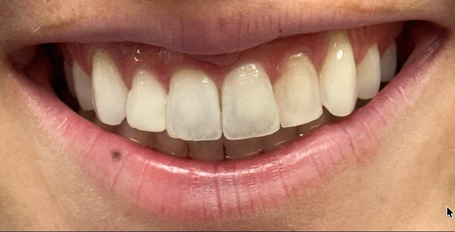
Fig 1
Chief Complaint:
The patient presented with concerns about the shape, symmetry, and color of her four upper front teeth. She noted that tooth #7 appeared shorter than tooth #10, which was longer, yellow, and discolored in comparison. She was also bothered by teeth #8 and #9, which showed a gray translucency that made them stand out from the rest of her smile. Additionally, she felt tooth #11 was longer and more pointed than tooth #6, contributing to an overall lack of symmetry across her smile.
Clinical Findings:
Clinical examination revealed existing composite restorations on the peg-shaped lateral incisors, #7 and #10, both showing visible discoloration. Teeth #8 and #9 exhibited gray translucency along the incisal half of each tooth. Incisal edge asymmetry was noted between #7 and #10, and #11 presented with a disproportionately long incisal edge relative to #6.
Diagnosis:
Tooth #7 is a peg lateral incisor with existing distofacial (DF) and mesiofacial (MF) composite bonding and is approximately 1mm short of ideal incisal length relative to #10. Teeth #8 and #9 both show excessive translucency in the incisal half, giving them a grayish appearance; #8 additionally presents with a tapered incisal edge contour. Tooth #10 is a peg lateral incisor with existing DF and MF composite filling that has discolored to a yellow shade over time, and is longer than #7. Tooth #11 has an elongated incisal edge that is noticeably prominent relative to the adjacent dentition.
Treatment Plan
A comprehensive case history was obtained, including medical and dental history, esthetic concerns, and patient expectations. Preoperative records included an initial intraoral scan used to generate a no-prep digital wax-up for the patient’s review.
Following the review of the initial digital wax-up with the patient, Dr. Frith performed minimal preparation on the case, reducing the aged composite build-up on peg laterals #7 and #10 so that over 90% of the resulting bonding surface on #10 would be natural tooth, lightly reducing the incisal edge of #7 to improve symmetry with #10, and further reducing the incisal edge of #11, which the patient felt was too long relative to #6. He then rescanned the patient to account for the prepared teeth and submitted new photography to guide the revised design.
Dr. Frith’s feedback for the updated design, based on the initial wax-up, included the following:
- Lengthen the incisal edge of #7 by approximately 0.5-1mm to better match #10.
- Preserve the incisal edge length of #6, which the patient liked, matching that proportion when finalizing the reduction already performed on #11.
- Account for the prepped-back composite on #7 and #10 following removal of the majority of the old peg lateral build-ups.
- Increase body opacity to eliminate the grayish appearance seen along the incisal half of the four front teeth in photos, without creating a flat, uniformly opaque “chicklet” appearance.
- Maintain the flat, even incisal edge line established in the initial mockup, having removed the angled appearance of the four incisal edges.
The finalized design of ultra-thin veneers for teeth #7-10 was fabricated at 100µm, calibrated to the post-preparation scan.
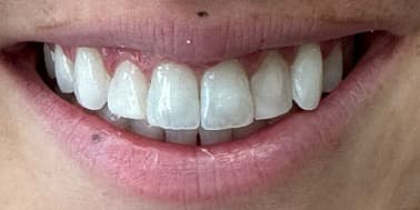
Fig 2
Fig 3
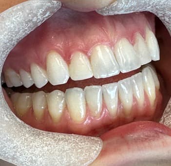
Fig 4
Fig 5
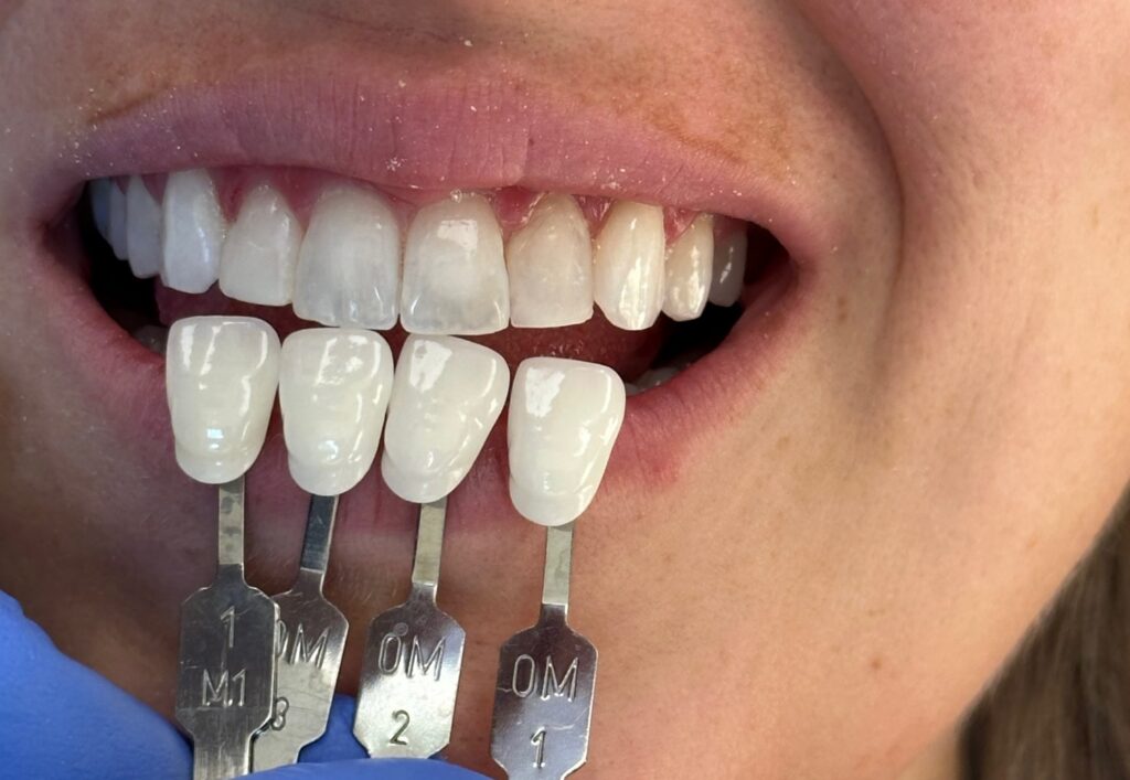
Fig 6
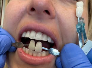
Fig 7
Fig 8
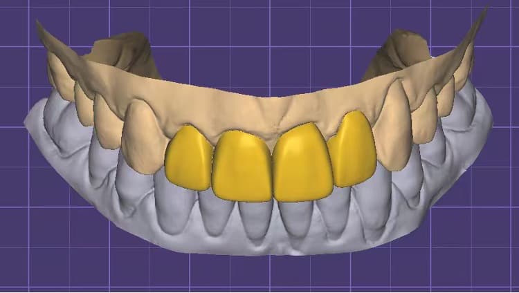
Fig 9
Fig 10
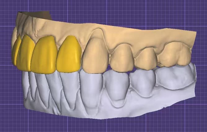
Fig 11
Fig 12
Treatment Procedure
The treatment began with a test fit of the veneers to evaluate fit and esthetics. Both a clear try-in paste and a white opaque try-in paste were used to determine the best cement shade for the case. Because the patient had recently whitened her natural teeth with dental bleaching trays, the clear try-in paste did not appear white enough against her natural shade, so the white opaque try-in paste was selected as the better match and used to guide the permanent cement shade. The veneers were then cleaned phosphoric acid for 30 seconds and then rinsed with water and air dried. Two layers of porcelain primer (silane) were brushed onto the veneers, allowed to sit for 5 minutes, then air dried for 30 seconds. A thin layer of porcelain bonding resin was applied without light curing. The veneers were preloaded with permanent Insure Universal White Opaque cement, matched to the white opacity shade confirmed during try-in, and protected under an orange shield after being warmed in the composite warmer. On the tooth surfaces, a micro air abrasion was used. The teeth were then etched with 32% phosphoric acid for 30 seconds, rinsed thoroughly for 30 seconds, and 1 to 2 thin layers of Bisco All-Bond Universal adhesive were applied, air thinned, and light cured for a minimum of 10 seconds at 800mW/cm2. The veneers were seated and tack-cured in place to stabilize positioning. Excess uncured cement was then carefully removed prior to final curing as cured cement is highly difficult to remove, especially interproximally. Once excess cement was removed, the veneers were light cured for 20 seconds. Interproximal saws and scalers were used to remove any residual cement. Contacts and occlusion were checked, and final radiographs were taken.
Outcome and Follow-Up
The four ultra-thin zirconia veneers were successfully placed on teeth #7-10, correcting the discoloration and shade mismatch from the aged composite build-ups, masking the gray incisal translucency of #8 and #9, and achieving improved symmetry between #7 and #10 and along the incisal edge line. The veneers fit precisely, with excellent marginal adaptation, and Dr. Frith felt all treatment goals had been achieved, a sentiment the patient shared. The outcome was beautiful, and both the patient and her mother were very pleased with the result. The patient loved looking at her new smile in the mirror and couldn’t stop smiling.
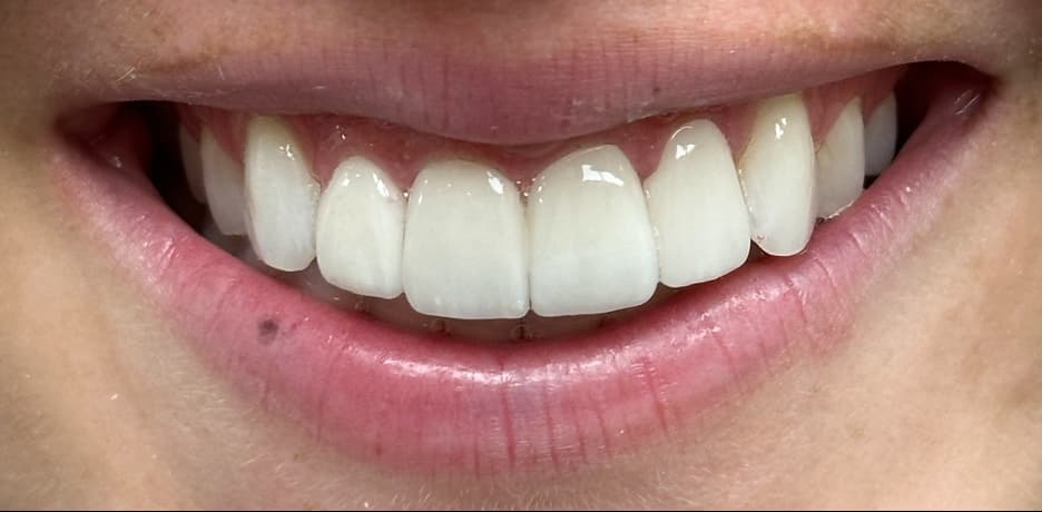
Fig 13
Discussion
This case illustrates the value of combining minimal preparation with an additive, ultra-thin veneer philosophy within a single treatment. Teeth #7 and #10, previously restored as peg laterals with composite that had discolored over time, required reduction of the existing material before a predictable bond and shade result could be achieved, while #8, #9, and #11 were addressed with a more conservative, largely additive approach focused on optical masking and minor incisal reshaping. The two-stage digital wax-up process, an initial no-prep version followed by a refined design based on the post-preparation scan, allowed the treatment plan to adapt once the actual amount of preparation needed on the peg laterals was known.
Achieving the patient’s desired 0M2 shade also required a deliberate balance between opacity and translucency. Increasing body opacity was necessary to mask the gray translucency along the incisal half of #8 and #9, but overcorrecting risked an unnaturally flat, uniform “chicklet” appearance. The final design aimed to preserve enough subtle translucency at the incisal edge to maintain a natural look while still eliminating the grayish cast the patient wanted addressed.
Conclusion
This case demonstrates that UltraThineer veneers can be adapted to cases requiring a mix of minimal preparation and additive correction, particularly when legacy composite restorations complicate an otherwise conservative treatment plan. By combining targeted preparation on teeth #7 and #10 with additive, prep-free design on #8 and #9, and using an iterative digital wax-up process informed by both dentist and patient feedback, this case achieved improved shade uniformity, symmetry, and a natural incisal appearance for teeth #7-10.
References
- Denry I, Kelly JR. State of the art of zirconia for dental applications. Dent Mater. 2008;24(3):299-307.
- Kelly JR, Denry I. Stabilized zirconia as a structural ceramic: an overview. Dent Mater. 2008;24(3):289–298.
- Magne P, Belser UC. Bonded porcelain restorations in the anterior dentition: a biomimetic approach. Chicago: Quintessence Publishing; 2002.
- Taraszkiewicz-Sulik K, Wiśniewski P, Cywoniuk E, Sierpińska T. Two-year clinical performance of ultra-thin no-prep veneers from 5Y-TZP zirconia: a retrospective study. Bioengineering. 2025;12(9):976.
- Alshehri A. Aesthetic treatment of bilateral peg-shaped lateral incisor and restoration of a harmonious smile: a case report. Case Rep Dent. 2025:3371424.
- Maroiu AC, et al. Spectrophotometric evaluation of the masking ability of 3D-printed ceramic-filled hybrid veneers on discolored dental substrates. Dent J (Basel). 2026;14(6):349.
- Coachman C, Calamita M. Digital smile design: a tool for treatment planning and communication in esthetic dentistry. QDT. 2012;35:1–9.